No products in the cart.



Have you ever been told that proteins are polar, only to learn that hydrophobic residues avoid water and drive folding? The apparent contradiction disappears once you stop treating polarity as a yes-or-no label. So, are proteins polar? The most accurate answer is that proteins are chemically mixed, spatially patterned, and strongly influenced by their environment.
A protein can present water-friendly regions at its surface while hiding water-avoiding regions inside its folded core. Its behavior also changes with amino-acid composition, folding, pH, charge state, solvent, and nearby surfaces. This guide follows that chemistry from individual amino acids to whole-protein behavior, then connects it to solubility, aggregation, binding, chromatography, and formulation.
The question sounds like a simple classification exercise, but it contains several questions. Are the amino acids polar? Does the protein carry charge in water? Which parts of the folded structure contact the solvent? How does the answer change when the buffer pH or surrounding surface changes?
Proteins aren't uniformly polar. A single chain may expose charged or hydrogen-bonding residues on one face while placing greasy, nonpolar residues inside a protected core. The result is often an amphipathic molecule, meaning it combines hydrophilic and hydrophobic regions rather than belonging completely to one category. The foundational relationship between amino-acid composition and protein behavior was recognized early in protein chemistry, including a 1965 Nature paper that described the ratio of polar to nonpolar amino acids as an important protein characteristic. That Nature paper remains useful background for understanding why composition matters.
A useful way to organize the topic is to separate four levels:
Students often search this question after a lecture has used “polar” and “nonpolar” as if they were permanent labels. That shortcut works for introducing side chains, but it becomes misleading for a folded protein. The practical outcome depends on the polarity profile across the molecular surface, not just on the average composition.
That profile influences whether a protein stays dissolved, associates with another molecule, sticks to a chromatography medium, or forms aggregates. It also helps explain why two proteins with similar size can behave very differently in the same buffer.
Practical rule: Ask which regions are exposed, which groups are charged, and what the surrounding solution is doing before assigning a protein a single polarity label.
What makes an amino acid polar, even when its overall charge is zero? The answer begins with its shared molecular framework: an alpha-amino group, an alpha-carboxyl group, and a variable side chain, often called the R group. After amino acids join through peptide bonds, the amino and carboxyl groups form the backbone, while the R group gives each residue much of its chemical character.
In water near physiological pH, around 7, all 20 standard amino acids predominantly occur as zwitterions. Their amino group carries a positive charge as an ammonium group, while their carboxyl group carries a negative charge as a carboxylate. A LibreTexts overview of amino-acid zwitterion forms explains this aqueous form and the amphiprotic behavior of amino acids.
A small magnet provides a useful comparison: it has two opposite poles even when the object as a whole is electrically neutral. In the same way, a zwitterion has positive and negative regions at once. Zero net charge does not mean zero polarity. Those separated charges create an uneven electrical environment and allow the molecule to interact with water.
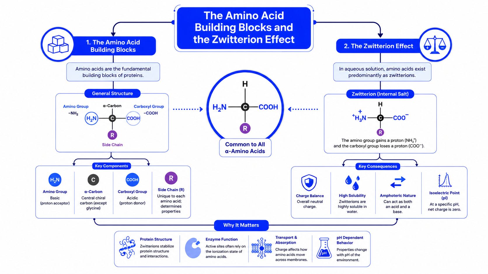
As amino acids form a protein, their amino and carboxyl groups become connected by amide bonds, also called peptide bonds. Atoms in these bonds can participate in hydrogen bonding, so the backbone contributes polar features even when a particular side chain avoids water.
The R groups create the larger pattern. Some contain hydroxyl or amide groups, others present hydrocarbon surfaces, and some gain or lose protons as conditions change. Along a folded protein, these groups form a surface pattern that influences water exposure, molecular binding, and the tendency to remain dissolved or aggregate.
For readers comparing protein chemistry with nutrition or laboratory materials, an amino acid supplement guide offers broader terminology. Free amino acids and amino-acid residues inside a folded protein are not interchangeable, because neighboring groups and the surrounding structure change their behavior.
Frederick Sanger's completion of the first protein amino-acid sequence, insulin, in 1949, helped establish that proteins are specific chains rather than uniform substances. The zwitterion overview connects this milestone with the modern view that protein chemistry arises from sequence, structure, and environment.
Are amino-acid residues polar or nonpolar? These categories help organize protein chemistry, yet they are not permanent labels. A residue's size, shape, charge, hydrogen-bonding ability, and ionization state all affect how it behaves within a folded protein.
Serine and threonine contain hydroxyl groups. These groups can form hydrogen bonds with water, so the residues are usually classified as polar and uncharged when their side chains carry no net charge. Asparagine and glutamine contain amide groups, which also interact with water through hydrogen bonding.
Cysteine and tyrosine show why the categories need context. Their sulfur- or hydroxyl-containing groups can take part in polar interactions, while the surrounding structure and protonation state influence their effective behavior. An amino-acid chemistry reference on polar and nonpolar side chains explains how electronegative atoms such as oxygen and nitrogen create uneven electron distribution that supports hydrogen bonding.
Leucine, isoleucine, and valine have largely hydrocarbon side chains. Phenylalanine adds an aromatic ring, and methionine contains sulfur within a mostly nonpolar structure. These residues generally present water-avoiding surfaces and often contribute to less water-exposed regions of a protein.
Hydrophobicity measurements at pH 7 show how widely residues can differ. Phenylalanine and isoleucine have normalized hydrophobicity values of 100 and 99, while serine and glycine are near the neutral end at -5 and 0, respectively, in the 1965 Nature hydrophobicity analysis of polar-to-nonpolar ratios. The scale describes residue behavior under defined conditions. It does not assign one polarity score to an entire protein, whose surface may contain alternating water-friendly and water-avoiding patches.
Lysine, arginine, and histidine are commonly described as positively charged, while aspartate and glutamate are commonly described as negatively charged. Their actual charge depends on pH and local chemical surroundings, particularly for groups near their ionization transitions. A pH shift can therefore change both electrostatic attraction and how readily a protein remains dissolved or binds another molecule.
| Category | Example residues | Key functional group |
|---|---|---|
| Polar, uncharged | Serine, threonine, asparagine, glutamine | Hydroxyl or amide |
| Nonpolar | Leucine, isoleucine, valine, phenylalanine, methionine | Hydrocarbon or largely nonpolar surface |
| Positively charged | Lysine, arginine, histidine | Basic, proton-accepting groups |
| Negatively charged | Aspartate, glutamate | Carboxylate groups |
These categories work as maps rather than fixed identities. A video introduction to amino-acid polarity can help visualize the functional groups, while protein behavior still depends on each residue's position, neighbors, and chemical environment.
A protein sequence is linear, but the molecule's chemistry is three-dimensional. Folding brings distant residues together, allowing the chain to form a compact structure in which water-friendly and water-avoiding groups occupy different locations.
Hydrophobic residues such as leucine, valine, and phenylalanine are often packed toward the interior, where they have less contact with water. Polar and charged residues such as serine, glutamate, and arginine are often more compatible with solvent exposure. Protein-structure teaching material describes this general arrangement, including the tendency for hydrophobic residues to occupy interiors and hydrophilic residues to contact aqueous surroundings.
The rule isn't absolute. Some nonpolar groups remain exposed because they participate in binding, membrane association, or protein-protein contacts. Some polar groups become buried when they form strong internal hydrogen bonds or salt bridges. Folding reflects the combined effect of many interactions, not one sorting instruction.
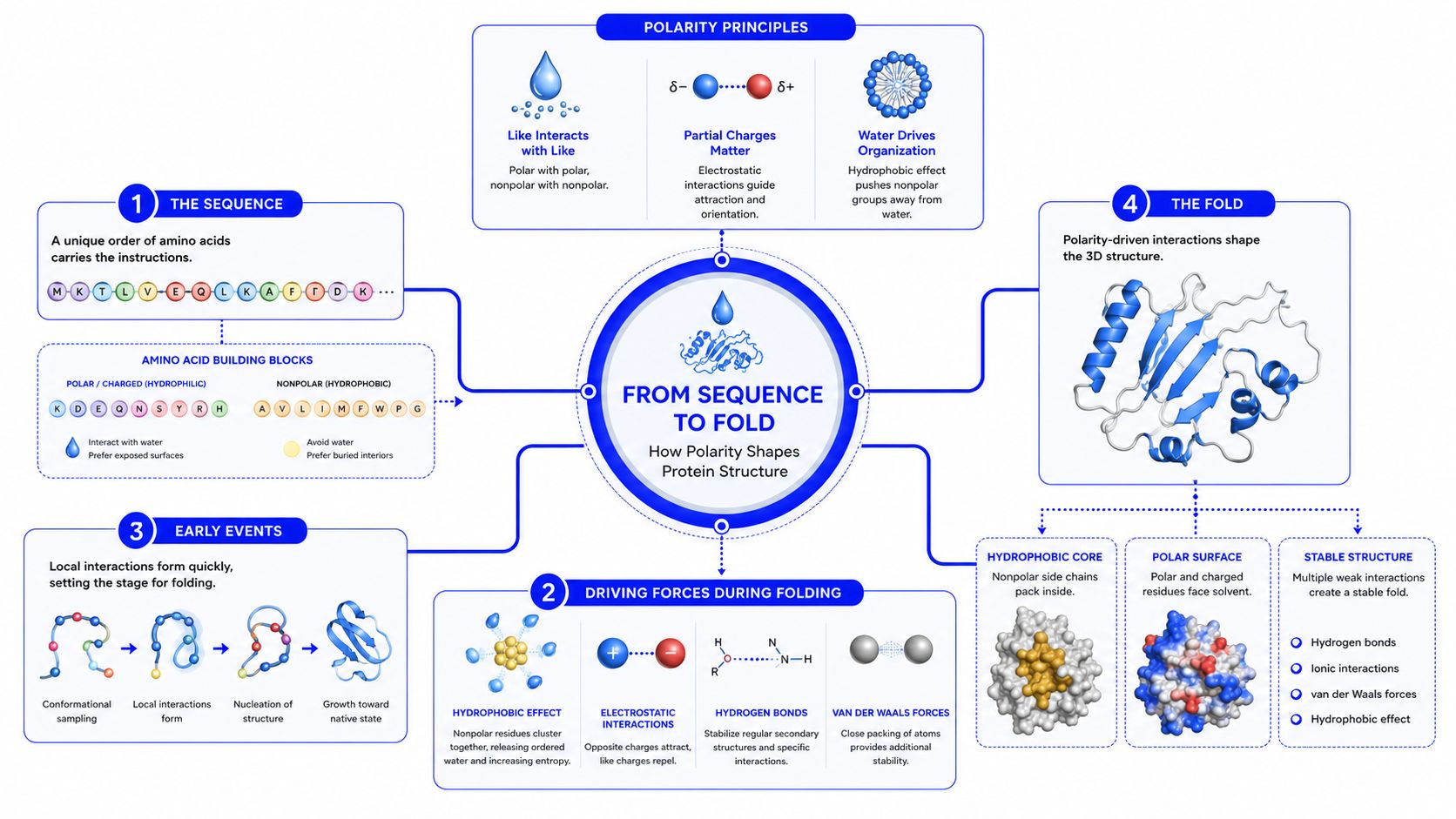
The backbone can form alpha helices and beta sheets through hydrogen bonding. Side-chain packing then stabilizes the larger fold, while the solvent-facing surface presents a patchwork of charged, polar, and nonpolar regions.
An amphipathic helix makes the pattern especially clear. One side can contain polar or charged residues that face water, while the opposite side contains nonpolar residues that face a membrane, another protein, or a protected interior. This arrangement gives the protein a directional surface rather than one uniform chemical character.
Surface patches also create recognition sites. A binding pocket may combine hydrogen-bond donors, acceptors, charged groups, and hydrophobic surfaces, allowing it to recognize one ligand more effectively than another. Protein surfaces aren't smooth chemical sheets. They're structured terrains, and their topology helps determine docking, membrane association, and selective binding.
A protein's sequence stays the same when you change a buffer, but the charge state of its ionizable groups can change. Protonation and deprotonation alter the electrical pattern on the surface, which changes how the protein interacts with water, salts, other proteins, and laboratory materials.
At approximately pH 7, the standard amino acids predominantly occupy zwitterionic forms, but side chains and terminal groups can respond differently to pH. The isoelectric point, or pI, is the pH at which a protein has no net charge. Below that point, the protein tends toward a more positive net charge; above it, the protein tends toward a more negative net charge.
That doesn't mean the molecule becomes chemically blank at its pI. Local positive and negative patches remain, even when the total charge sums to zero. This is why proteins can still bind surfaces or aggregate near conditions where their net charge is minimal.
| Residue | Typical pKa | Dominant form near pH 7 | Charge state |
|---|---|---|---|
| Aspartate | About 4 | Deprotonated carboxylate | Negative |
| Glutamate | About 4 | Deprotonated carboxylate | Negative |
| Histidine | About 6 | Often neutral, with a fraction protonated | Environment-dependent |
| Lysine | About 10 | Protonated amino group | Positive |
| Arginine | About 12 | Protonated guanidinium group | Positive |
These values are approximate chemical reference points, and local structure can shift a group's behavior. The MCAT amino-acids and proteins guide provides useful context for pH-dependent charge and the role of the isoelectric point.
Ionic strength can shield electrostatic attractions and repulsions. A co-solvent such as glycerol or ethanol can change how water organizes around exposed groups, while denaturants such as urea can disrupt the interactions that support a native fold. Temperature also changes molecular motion and solvent interactions.
In practice, researchers may observe changes in solubility, viscosity, or aggregation during storage and assay setup. A protein that remains clear in one buffer can become cloudy in another because the surface charge pattern, hydration layer, or folded state has changed.
Lab interpretation: A change in apparent polarity often reflects the solution and the protein's conformation together, not a change in the amino-acid sequence.
Protein polarity becomes operationally important as soon as a researcher tries to dissolve, stabilize, purify, or compare a sample. Exposed polar and charged groups generally support hydration, while exposed hydrophobic patches can encourage protein-protein association. Those associations may produce aggregation or, during expression, contribute to insoluble inclusion bodies.
Researchers can sometimes alter the surface character of a construct with a fusion tag or a modification such as PEGylation. These approaches don't erase the underlying sequence, but they can change the accessible interface and improve compatibility with the chosen solution. Any such strategy still requires experimental validation because a modification can also affect folding or binding.
Different chromatography modes emphasize different interactions:
The choice of method depends on the molecule and the desired state. A comparison of preparative HPLC and SFC notes that preparative HPLC can handle a broad range of sample types, including large proteins, while SFC can be less suitable for highly polar compounds and protein separations.
A formulation scientist may adjust buffer pH, salt type, surfactants, or stabilizers to control effective surface interactions. The goal might be to keep a protein hydrated, reduce unwanted adsorption, limit aggregation, or preserve the fold during shipping and storage.
The right condition isn't determined by polarity alone. Researchers must also consider the protein's pI, binding requirements, concentration, temperature, container surface, and intended assay. A successful formulation balances those variables rather than maximizing one type of interaction.
The first misconception is that every protein dissolves equally well in water. Many soluble proteins expose substantial polar and charged surface area, but membrane proteins often contain hydrophobic regions designed to contact lipid environments. Fibrous proteins can also have repeating structural patterns that don't behave like compact globular enzymes, and proteins with exposed hydrophobic patches may aggregate or require detergents.
The second misconception is that molecular size predicts polarity-driven behavior. A small peptide can contain a strongly polar sequence and interact readily with water, while a larger multimeric enzyme can bury many nonpolar surfaces inside its structure or at subunit interfaces. Size influences diffusion and physical handling, but it doesn't tell you which chemical groups are exposed.
A further mistake is to treat polarity as an average property stamped across the sequence. Researchers often need to know the polarity of a particular surface region, binding pocket, interface, or membrane-facing segment. Research on protein surface hydration and polarity supports the importance of spatially distributed polar and nonpolar surface domains.
Misreading surface chemistry can lead a team to choose an unsuitable buffer, expect solubility that the protein can't provide, or interpret nonspecific binding as a specific interaction. It can also produce a failed purification when the selected chromatography mode doesn't match the accessible charge or hydrophobicity profile.
A better mental model is a patchwork map. Each region contributes differently, and the map can shift as the protein folds, associates, unfolds, or enters a new chemical environment.
So, are proteins polar? They're neither uniformly polar nor uniformly nonpolar. Each residue contributes a distinct chemical character, the backbone supplies hydrogen-bonding capacity, folding often buries hydrophobic side chains, and the exposed surface presents a mixture of polar, charged, and nonpolar patches.
The environment then changes the apparent behavior. pH redistributes charge, ionic strength modifies electrostatic interactions, solvents alter hydration, and crowding or nearby surfaces can favor association. That's why the same sequence may show different solubility, binding, or aggregation behavior in different buffers.
Keep these principles in mind when planning an experiment:
Researchers studying peptides and proteins can build on these principles through peptide synthesis resources, protein-expression guides, and solubility-troubleshooting material. The benefits of peptides and the usefulness of related compounds depend heavily on sequence, purity, folding, formulation, and the intended research context, so chemical behavior should guide experimental design rather than marketing labels.
Understanding polarity also makes analytical results easier to interpret. A shift in retention, precipitation, or binding may be telling you that the accessible surface has changed, even when the molecular identity appears unchanged.
Peptide Warehouse USA offers research peptides and related compounds for laboratory, analytical, and preclinical applications, with batch documentation designed to support traceability and informed selection. Visit Peptide Warehouse USA to explore options and review research-use materials that can help you plan experiments around peptide sequence, purity, and solubility.