No products in the cart.



Most therapeutic peptides clear the bloodstream in minutes to a few hours, but biological effects and detectability can extend far longer than that single half-life figure suggests. Some short-acting peptides may leave circulation in 12–24 hours, while metabolites can remain detectable for 24–48 hours or longer, and modified analogs may persist for days to weeks.
So, how long do peptides stay in your system? The honest answer depends on what you mean by “stay.” Are you asking how long the molecule remains in blood, how long it remains in tissues, how long it continues producing a biological response, or how long a laboratory can detect the parent compound or its metabolites?
Most online explanations offer one half-life number and stop there. That shortcut creates the central misunderstanding. A peptide can fall below a blood test's detection threshold while downstream signaling continues, or a urine test can identify a metabolite after the original compound is no longer pharmacologically active.
This guide separates those timelines into three practical lenses: blood presence, tissue residence, and detectability. It also explains why renal clearance, molecular design, administration route, assay sensitivity, and receptor biology can all change the answer.
The phrase “stay in your system” combines several different questions that need separate answers.
This refers to the amount of intact peptide present in plasma or blood. For many therapeutic peptides, that period is relatively short because the kidneys filter small, hydrophilic molecules and enzymes break peptide bonds. The measurable concentration can fall quickly even when the body's response doesn't stop at the same moment.
Blood isn't the only compartment that matters. A molecule may distribute into tissues, bind to albumin, attach to a receptor, remain near an injection site, or move more slowly from a formulation designed for extended release. Tissue exposure can therefore outlast the period when a routine blood sample shows much of the parent peptide.
Detection is its own clock. An assay may identify the parent peptide, a metabolite, an immunoreactive fragment, or another marker. The result depends on the sample type, collection time, analytical method, cutoff concentration, and the specific molecule being measured.
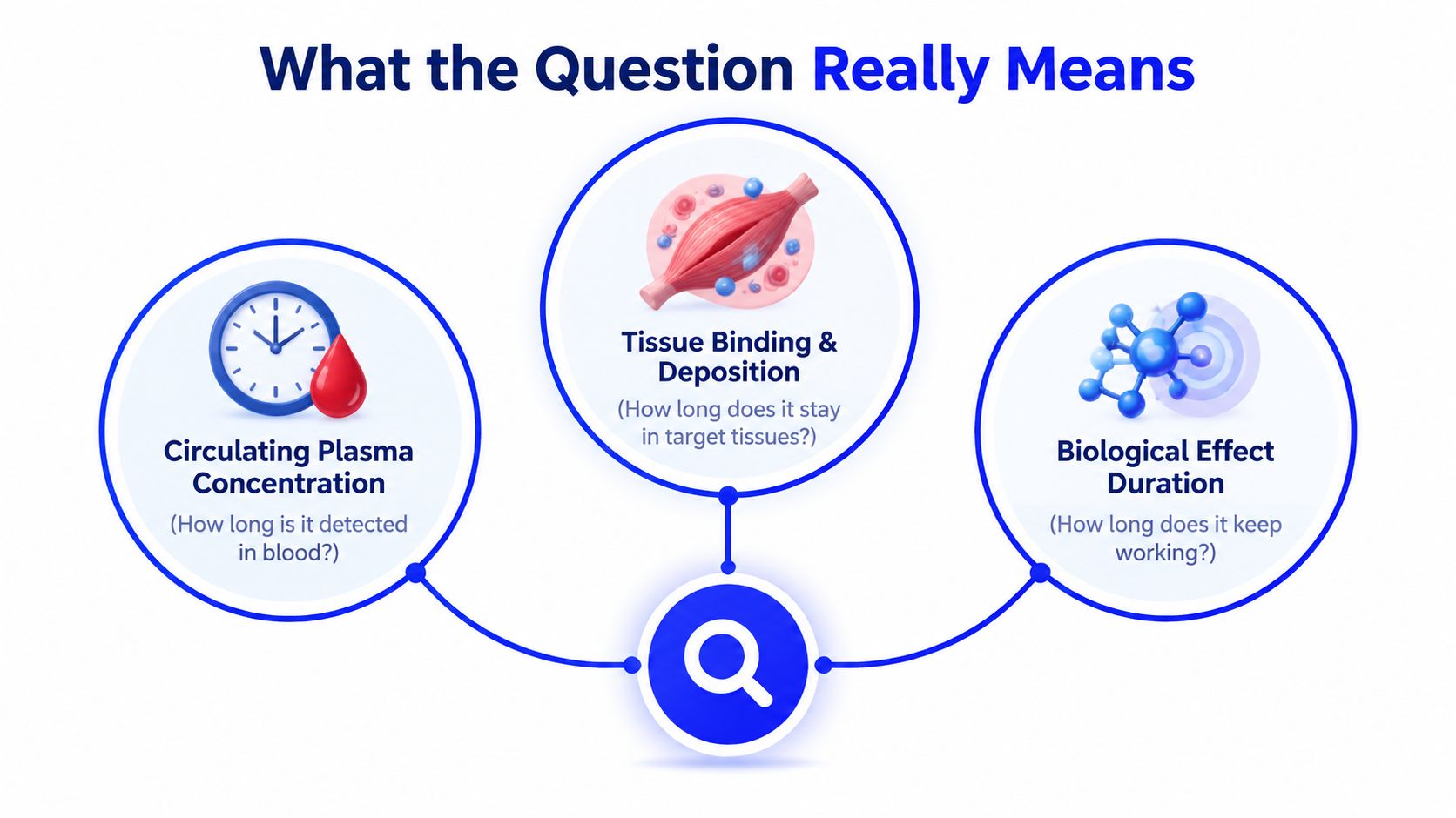
A useful mental model is:
The mistake is treating “undetectable in blood,” “no longer biologically active,” and “completely eliminated” as interchangeable. They aren't. The sections that follow examine each layer separately, using concrete peptide examples and the pharmacokinetic principles that explain why timelines vary.
How can a peptide leave the bloodstream quickly yet remain relevant in the body? Three terms help answer that question: half-life, clearance, and volume of distribution. They describe different parts of the same process, so none should be treated as the exact time until a peptide is gone.
A peptide's elimination half-life is the time required for its plasma concentration to fall by half. A bathtub with colored water draining provides a useful comparison. The color becomes less intense as water leaves, but the drain does not remove every colored molecule at once.
After one half-life, about half the measured amount remains. After another, about half of that remainder remains. Researchers commonly use approximately 4–5 half-lives as the point when most of a compound has cleared, or roughly 97 percent clearance in the simplified model described in peptide pharmacokinetic literature (Frontiers review of therapeutic peptide delivery).
Clearance is the body's capacity to remove a peptide from plasma, often reported in units such as mL/min/kg. Kidney filtration may contribute substantially. Enzymatic breakdown, liver processing, cellular uptake, and other pathways can also reduce the amount circulating.
Clearance answers a different question from half-life. It describes how efficiently the body removes the peptide, while half-life describes how quickly the measured concentration falls under particular conditions.
Volume of distribution, or Vd, is an apparent measure of how widely a peptide spreads beyond the blood. A small, water-loving peptide may stay mainly in plasma before removal. A peptide that enters tissues or binds albumin can show a larger apparent volume.
Half-life depends on both clearance and volume of distribution. A peptide may be removed efficiently from plasma yet show a longer terminal decline if it distributes into tissues and returns gradually to the bloodstream. That return can make the concentration curve look like a slow second phase rather than a simple drop.
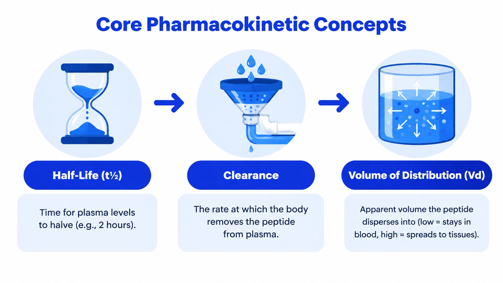
Steady state is generally reached after about 4–5 half-lives, rather than after one dose or a fixed calendar interval. A half-life helps estimate plasma concentration decline. It does not by itself establish how long tissue effects continue, how long a laboratory can detect related material, or when all biological activity has ended.
Native peptides often disappear from circulating blood quickly because the body has several overlapping ways to remove them. Small molecules can be filtered by the kidneys, while enzymes can break their amino-acid chains into smaller fragments.
The kidney filters small, water-soluble peptides through the glomerulus. Peptides below the roughly 60-kDa filtration boundary can be cleared particularly readily because their size and hydrophilicity make them suitable for renal handling, as discussed in research on kidney clearance and peptide drug design.
After filtration, a peptide may be reabsorbed and degraded by proximal tubule cells, or it may move toward urinary excretion. This is one reason many unmodified peptides have plasma half-lives measured in minutes rather than days.
Peptidases in blood and tissues can cleave peptide bonds. A molecule doesn't need to be physically excreted as an intact chain before its original pharmacologic identity is lost. Once enzymatic cleavage changes the structure, an assay may no longer recognize the parent compound, and the fragment may not interact with the same receptor.
The liver can metabolize some peptide structures, while receptor-mediated endocytosis can pull a peptide into cells after it binds its target. Distribution into tissues can also alter the apparent decline in plasma.
| Clearance Route | Mechanism | Example Peptides | Typical Clearance Time |
|---|---|---|---|
| Renal filtration | Small, hydrophilic peptides pass through the glomerular filter and undergo reabsorption or excretion | Many native therapeutic peptides | Minutes to hours |
| Proteolytic degradation | Blood and tissue peptidases cleave peptide bonds | Short-lived signaling peptides | Minutes to hours |
| Hepatic handling | Liver enzymes and cellular processes alter or remove peptide structures | Molecule-dependent | Varies by structure |
| Receptor-mediated uptake | Target cells internalize bound peptide and process it | Receptor-active therapeutic peptides | Varies by receptor and tissue |
These mechanisms exist because peptides are biological signaling molecules, and the body needs to regulate them rather than allow every signal to remain indefinitely. The resulting short blood residence doesn't automatically predict a short-lived physiological response.
How long a peptide remains in the body depends on its sequence, formulation, administration route, assay, and molecular modifications. The ranges below are useful comparisons, not fixed promises for every product in a category. They describe plasma half-life, the decline of parent peptide in circulation, rather than the full duration of tissue exposure or biological effects.
Native signaling peptides often disappear from plasma quickly. Oxytocin may have a half-life of 1–6 minutes, vasopressin analogs 10–35 minutes, calcitonin about 1 hour, and insulin analogs 1–6 hours, with formulation affecting the result.
| Peptide family or format | Illustrative half-life | Main reason for duration |
|---|---|---|
| Oxytocin | 1–6 minutes | Rapid enzymatic and renal clearance |
| Vasopressin analogs | 10–35 minutes | Structure-dependent metabolism and clearance |
| Calcitonin | About 1 hour | Native peptide handling and degradation |
| Insulin analogs | 1–6 hours | Formulation and analog design |
| Short-acting native peptides | Minutes to a few hours | Limited protection from peptidases and filtration |
| Modified long-acting constructs | Hours to days or weeks | Albumin binding, larger carrier structures, or protease resistance |
Chemical design can move a peptide into a different duration category. Lipidation can promote albumin binding, allowing some medicines to circulate longer. PEGylation adds a hydrophilic protective layer that can reduce rapid enzymatic or renal removal. Fc-fusion attaches the peptide to a larger carrier, changing its distribution and recycling behavior. These approaches help explain why modified peptides may outlast native versions by days or weeks.
Half-life answers one narrow question: how quickly plasma concentration falls by half. It does not establish when receptor interaction ends, when a downstream biomarker returns to baseline, or when a laboratory can no longer identify a metabolite. Plasma clearance, tissue residence, and detection windows are related, but they are separate timelines.
Readers examining a long-acting GLP-1 medicine may find this Mounjaro timeline with Trim useful for distinguishing treatment-response timing from molecular clearance. Keep those categories separate when interpreting how long a peptide “lasts.”
A blood concentration curve and a biological-effect curve can look different. The first tracks how much parent peptide remains in circulation. The second tracks what happens after the peptide reaches a target, binds a receptor, activates signaling, and triggers a measurable response.
A peptide may fall below a blood assay's detection limit while some molecules remain bound to receptors or distributed in extracellular spaces. A subcutaneous formulation may also release material gradually from an injection site rather than sending the entire amount into circulation immediately.
The reverse can happen too. A laboratory may detect a small amount of parent peptide in plasma, but the concentration may be below the level needed to produce a sustained effect. Detectable doesn't necessarily mean functionally active, and undetectable in one blood sample doesn't prove that every biological consequence has ended.
The duration of an effect depends on several features:
Insulin is a familiar example of why plasma half-life and biological action should not be treated as identical. Its circulating half-life is short, yet glucose-related effects reflect distribution, receptor engagement, and downstream physiology rather than only the time intact insulin remains in blood.
For readers comparing scientific context with clinical discussions, resources such as therapeutic peptides at Aspire for Wellness can provide a separate overview of how providers frame peptide-related treatment questions. Any individual use, however, requires appropriate professional evaluation rather than inference from a half-life table.
A detection window answers a laboratory question, not necessarily a pharmacology question. Testing asks whether a method can identify a compound, fragment, or metabolite in a specific sample. Half-life asks how the parent concentration declines, usually in plasma.
Blood tests often target the parent peptide while its concentration remains above the assay's detection threshold. The window can be short when the molecule clears rapidly or when the test requires a highly specific intact structure.
Urine can tell a different story. The kidneys may excrete intact material or metabolites after plasma concentration has become very low. Hydration, kidney function, collection time, cutoff concentration, and assay design all influence the result.
| Concept | What it measures | Key determinants |
|---|---|---|
| Plasma half-life | Decline of parent peptide concentration in blood | Clearance, distribution, formulation, route |
| Blood detectability | Whether an assay identifies the target in plasma | Detection threshold, specificity, timing |
| Urine detectability | Parent compound or metabolite excretion | Renal handling, hydration, metabolism, sample timing |
| Pharmacologic activity | Whether enough active material or downstream signaling remains | Receptor interaction, tissue exposure, biological pathway |
| Mass-spectrometry signal | A specific molecular structure or metabolite | Instrument sensitivity, target selection, sample preparation |
Independent information on peptide testing notes that some short-acting peptides may leave circulation within 12–24 hours, while advanced mass spectrometry may detect metabolites for 24–48 hours or longer (peptide detectability and drug testing overview). Longer-acting analogs may remain relevant for days or weeks, depending on the molecule and its design.
A specific example illustrates the mismatch. Testing references report GHRP-2 metabolites detectable for up to 47 hours after administration, while some metabolite-based urine methods extend detection to about 4 days (peptide drug-testing guide). Those figures describe analytical findings under particular methods. They do not mean the parent peptide remained equally active for that entire period.
Practical rule: A negative blood result, a positive urine result, and a continuing biological effect can all occur at different points in the same timeline.
Immunoassays may recognize immunoreactive fragments or related structures, whereas mass spectrometry can target a more specific parent compound or metabolite. That is why a test result needs interpretation in the context of the molecule, assay, route, dose, sample type, and collection time.
Molecular design often determines whether a peptide behaves like a short-lived signal or a longer-acting therapeutic construct. Administration and patient-specific variables then shape the observed timeline.
Fatty-acid conjugation can encourage albumin binding. Albumin acts as a circulating carrier, which can reduce rapid filtration and protect part of the molecule from immediate enzymatic breakdown. Long-acting GLP-1 designs use this general strategy to extend exposure.
PEGylation attaches polyethylene glycol chains that increase hydrodynamic size and create steric protection around the peptide. The result can be slower proteolysis and reduced glomerular clearance, although the exact effect depends on the construct.
Fc fusion attaches the peptide to an antibody-derived Fc region. The larger format changes distribution and can take advantage of recycling pathways involving the neonatal Fc receptor.
Other sequence changes can make peptide bonds harder for enzymes to cut. D-amino-acid substitutions, N-methyl substitutions, cyclization, and related design choices may improve proteolytic stability, but each can also affect receptor binding and biological activity.
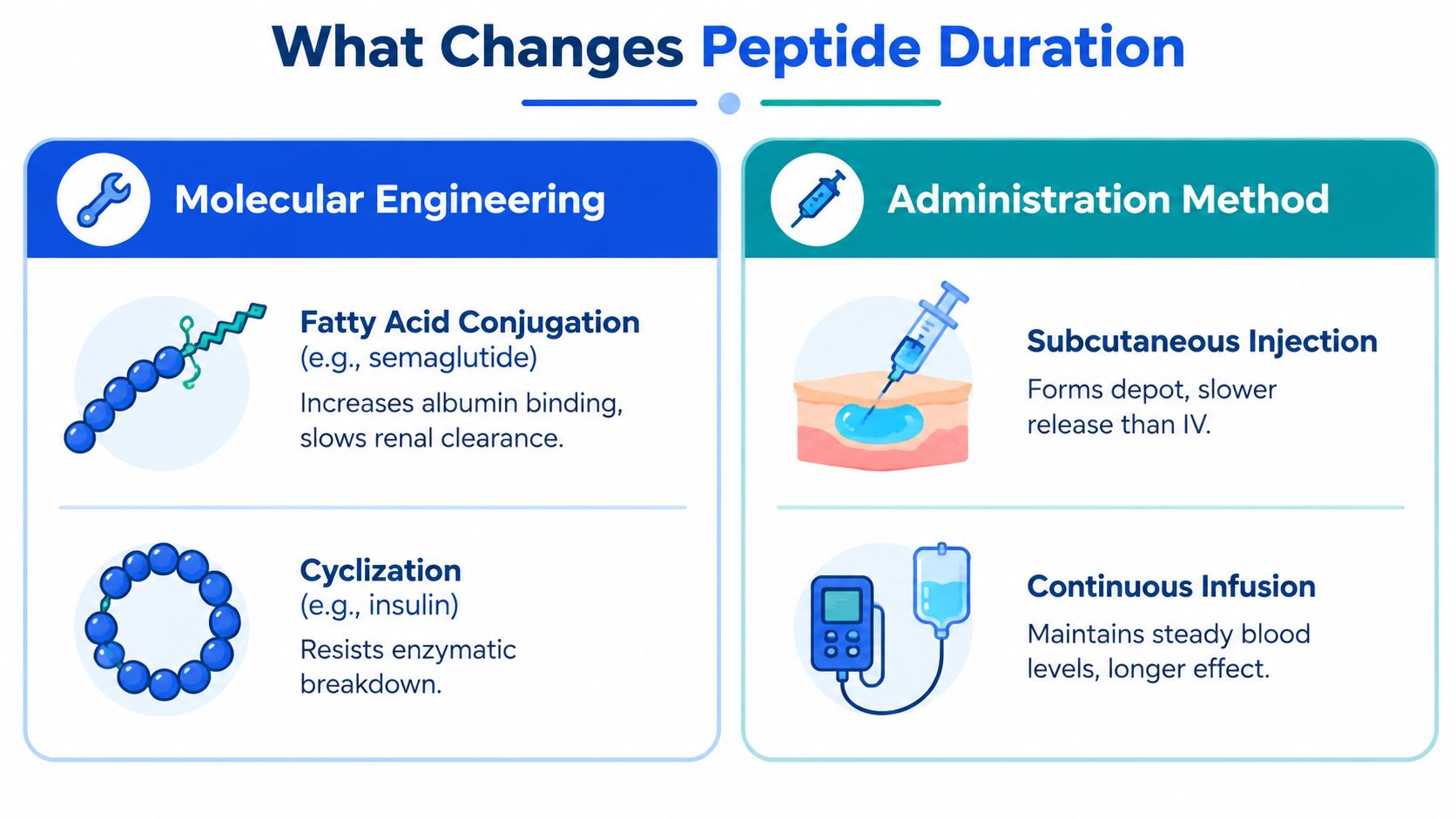
Intravenous administration places a peptide directly into circulation, while subcutaneous administration can create an absorption phase and a local depot. Sustained-release microspheres can slow release further. A continuous infusion can maintain a steadier plasma input compared with intermittent bolus dosing.
Dose frequency matters because repeated doses can overlap before the previous dose has fully declined. This is why steady-state interpretation requires the half-life rather than relying only on the calendar interval.
Kidney function, liver handling, body composition, injection site, concurrent medications, and the exact formulation can all influence peptide exposure. Literature values are averages from defined study conditions, not guarantees for every person or every research preparation.
For investigators trying to understand how measurements translate into formal interpretation, this resource on clarifying drug levels in Texas death investigations offers useful context on why laboratory concentration data must be considered alongside sampling conditions and analytical limits.
How long a peptide stays in the body depends on which endpoint you measure. A useful research model separates three questions:
Choose the endpoint before setting the timeline. A plasma pharmacokinetic study requires blood samples timed around absorption, peak concentration, and elimination. A tissue-distribution study may require tissue analysis or a validated surrogate. A testing study may rely on urine collection and an assay designed for the expected metabolite, rather than the intact parent molecule.
Pooling unrelated peptides into one “average half-life” analysis can hide major differences. A rapidly filtered native peptide and a longer-lasting modified analog may share a therapeutic category while showing different plasma, tissue, biological, and detection profiles.
For elimination studies, researchers should plan sampling across at least three to five half-lives. This range helps capture the decline toward low concentrations, although it does not replace molecule-specific validation or account for tissue residence and delayed effects.
The central lesson is simple: there is no single answer to how long a peptide stays in your system. Plasma clearance, tissue exposure, biological response, and laboratory detection describe related but distinct timelines.
Peptide Warehouse USA offers research peptides and related compounds for laboratory, analytical, and preclinical applications, with batch documentation such as Certificates of Analysis and stated purity information. Visit Peptide Warehouse USA to review research-use options and product documentation before planning work involving peptide clearance, stability, or detection.