
No products in the cart.



What happens when you add a “growth” factor to a cell culture? Do the cells multiply faster, or can the same molecule make them survive, migrate, differentiate, or even respond less effectively at a higher dose? That distinction is central to understanding a cell growth factor.
Growth factors are not interchangeable healing boosters or universal switches. They are signaling molecules whose effects depend on receptor expression, cell identity, ligand availability, timing, assay design, and the surrounding biological environment. This guide explains how growth-factor signaling works, how researchers measure it, why concentration alone can mislead, and how to evaluate recombinant proteins, peptide-related compounds, and other research materials with greater discipline.
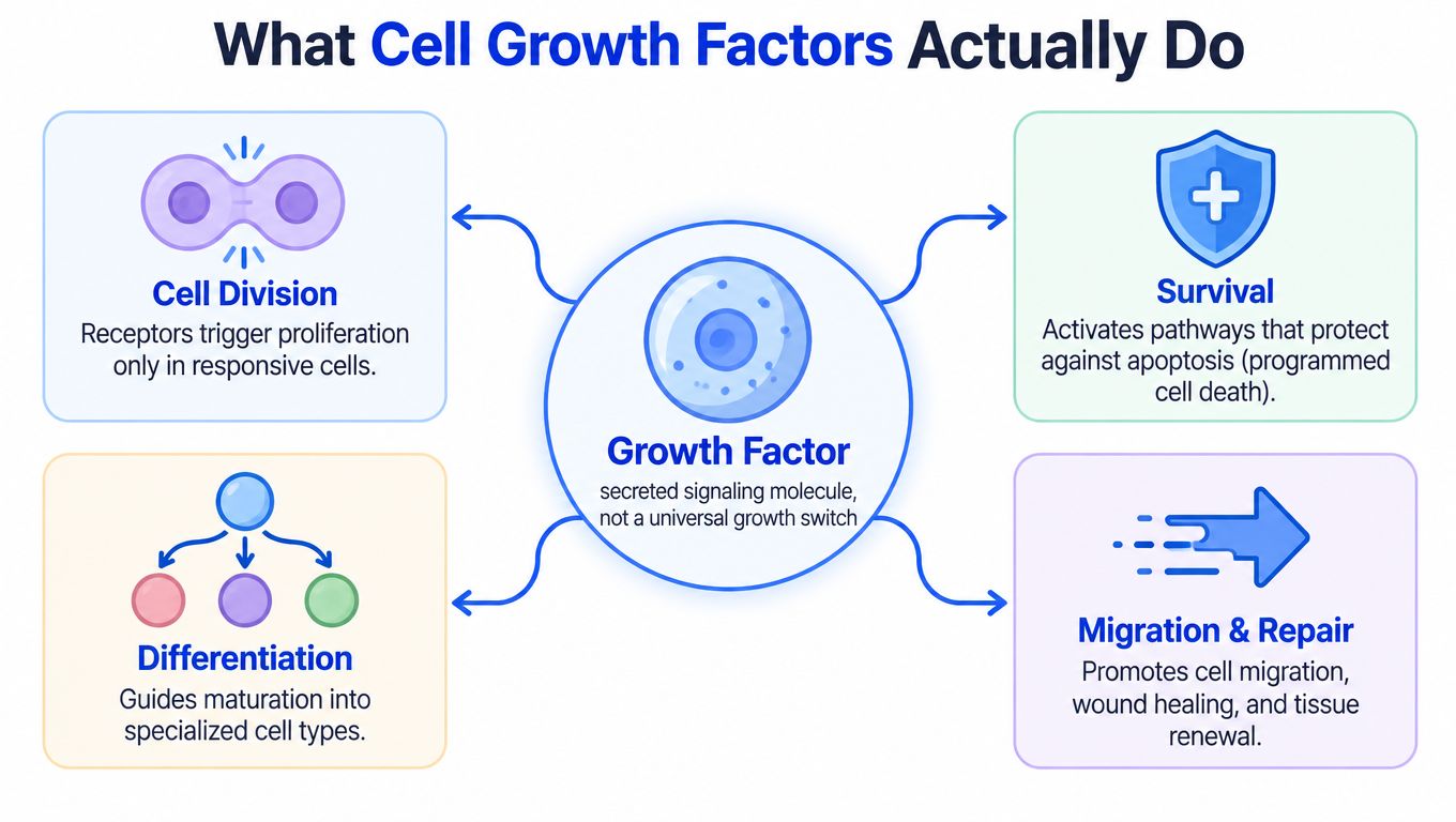
A cell growth factor is a secreted, biologically active molecule that helps regulate whether a cell grows, divides, survives, differentiates, or migrates. The term describes a broad class of signaling proteins, not one product or one biological action. A factor can support proliferation in one responsive cell model while producing a different outcome in another model.
The first step is receptor recognition. A growth factor binds to a matching cell-surface receptor, often a receptor tyrosine kinase, and that interaction starts intracellular signaling cascades. The resulting signals can change gene expression, promote the synthesis and accumulation of proteins and other macromolecules, and reduce their degradation. These processes increase cell mass, which is different from proliferation. Growth means increased cell mass, while proliferation requires division into new cells. Research on the biology of cell growth factors describes this distinction and the receptor-driven mechanism in detail.
Researchers commonly study purified proteins, recombinant factors, peptide-related signaling compounds, and cell-culture supplements to examine these outcomes. The resulting observations describe biological or preclinical research. They aren't guarantees of an outcome in humans, and they shouldn't be interpreted without the cell model, assay conditions, and reagent documentation.
The same principle applies to tissue-repair discussions. A growth factor may influence migration or cell survival, but the surrounding environment still matters. For readers interested in how local moisture conditions can affect repair environments, this overview of why moisture speeds recovery offers useful context without replacing controlled laboratory evidence.
Growth-factor signaling follows a sequence, but the sequence branches quickly. Consider fibroblast growth factors, or FGFs. Ligand binding activates one of four conserved transmembrane receptor tyrosine kinases, known as FGFR1, FGFR2, FGFR3, and FGFR4. The receptors then associate as a dimer, and their intracellular kinase domains phosphorylate one another on tyrosine residues. The documented FGF and FGFR signaling mechanism describes how these phosphorylation sites recruit downstream signaling proteins.
Those docking sites don't lead to one simple “growth pathway.” FGF signaling can engage several routes:
The balance among these routes helps explain why identical nominal treatments can produce different results. A cell with abundant receptors may respond strongly, while another cell with limited receptor expression may show little response. Adaptor proteins, cell state, feedback loops, exposure duration, and other ligands can further alter the outcome.
Insulin-like growth factor 1, or IGF-1, acts through the IGF-1 receptor, a receptor tyrosine kinase related to the insulin receptor. Its signaling can engage RAS-MAPK, PI3K-AKT, and JAK-STAT pathways. IGF activity is also moderated by insulin-like growth-factor-binding proteins, which bind and transport IGF ligands in extracellular fluids and circulation. That binding layer means the nominal amount of IGF-1 in a system doesn't necessarily equal the amount available for receptor engagement.
For this reason, cell number alone is a weak endpoint. A culture may eventually contain more cells, but that result doesn't show when signaling began or which pathway responded first. Measuring phospho-ERK and phospho-AKT over time can help distinguish early pathway engagement from later changes in proliferation. The NCI description of growth-factor biology emphasizes that these pathways are interconnected, especially through Ras-Raf-MEK-ERK and PI3K-AKT, rather than operating as one linear chain. The NCI growth-factor concept report provides the relevant signaling framework.
A factor's name tells you what molecule you used. It doesn't tell you what the molecule will do in your assay.
PDGF is a foundational example because it was identified as one of the earliest mitogens, meaning a signal capable of stimulating cell division. EGF demonstrates the wider range of growth-factor activity. It can promote proliferation, differentiation, growth, migration, and survival while inhibiting apoptosis. Those effects still depend on receptor abundance, cell type, exposure time, dose, and the other signals present in the culture.
The experimental context also determines how a result should be interpreted. An increase in cell number could reflect proliferation, improved survival, altered attachment, or a combination of processes. A migration assay may show movement without demonstrating durable tissue organization. A pathway assay may show receptor engagement without proving a later functional benefit.
| Growth factor | Typical receptor family | Common research context |
|---|---|---|
| EGF | EGFR, a receptor tyrosine kinase | Epithelial signaling, proliferation, migration, and survival |
| PDGF | PDGF receptors, receptor tyrosine kinases | Mitogenic signaling, stromal-cell behavior, and repair models |
| FGF | FGFR1, FGFR2, FGFR3, or FGFR4 | Cell-cycle signaling, differentiation, migration, and tissue models |
| IGF-1 | IGF-1 receptor, a receptor tyrosine kinase | Survival, metabolism, proliferation, and growth-factor regulation |
| VEGF | VEGF receptor family | Endothelial signaling and vascularization research |
| TGF-β, BMP | Serine-threonine kinase receptor systems | Differentiation, matrix biology, and remodeling research |
A useful comparison framework asks four questions:
Dysregulated growth-factor signaling is associated with tumor growth, angiogenesis, and therapeutic resistance. The association doesn't mean every laboratory use creates those outcomes, but it does show why researchers must treat signaling as context-dependent rather than automatically beneficial. A stronger signal isn't always a better result.
The right assay depends on the question. If you're asking whether a treatment changes cell number, use a proliferation readout. If you're asking whether the factor activates a receptor pathway, measure a signaling marker. These questions overlap, but they aren't identical.
A proliferation assay can estimate changes in cell number or DNA synthesis, but it may not separate faster division from improved survival. A migration assay can capture movement through a defined test environment, yet it doesn't establish that cells formed functional tissue. A differentiation assay can identify changes in markers or morphology, but marker expression alone may not prove mature function.
Pathway-read assays add an earlier layer of evidence. Measuring phospho-ERK or phospho-AKT at multiple time points can show whether receptor signaling was engaged before a later proliferation change appears. A credible design often combines an early pathway readout with a later cell-behavior endpoint instead of relying on cell count alone.
A concentration such as nanograms per milliliter describes the medium. It doesn't necessarily describe the effective exposure per cell. In an IGF-1 study using OVCAR5 ovarian-cancer cells, proliferative response was predicted more reliably by the total ligand available per cell than by medium concentration alone. Changing cell density altered the response even when the applied concentration stayed the same. The IGF-1 dose-per-cell study supports recording viable-cell count, seeding density, medium volume, and ligand mass.
A practical dose-response record should include:
Higher dosing also isn't more stimulatory. In chicken granulosa cells, FGF-2 maximized DNA synthesis at 0.1 ng/mL, while the maximum in theca cells occurred at 0.5 ng/mL. Doses above 25 ng/mL reduced synthesis to near-baseline levels in that study. These values are model-specific, not universal recommendations, and they illustrate why a complete response curve matters more than a single high-dose comparison.
A growth-factor reagent is only useful when the laboratory can connect its identity and handling history to the observed result. Before comparing products or batches, record the protein or peptide name, sequence or identity information where available, stated purity, lot number, preparation details, and assay conditions. A Certificate of Analysis, microbial report, endotoxin report, and potency information can help researchers judge whether the material is appropriate for a specific experiment.

Follow the supplier's documented storage and reconstitution instructions rather than assuming that every growth factor behaves the same way. Keep preparation records with the date, operator, solvent or buffer, working concentration, aliquot identity, and any deviation from the stated protocol.
A practical checklist includes:
Avoid treating nominal purity as a complete description of biological performance. Receptor context and assay conditions still determine activity, and a highly purified material can produce a weak result if the model lacks the relevant receptor or if the endpoint is poorly matched.
The following video can serve as general visual context for laboratory handling and research workflows:
Research materials may include purified proteins, recombinant factors, peptide-related compounds, or culture supplements. These materials support laboratory, analytical, and preclinical work, not guaranteed human outcomes. The NCBI overview of growth factors reinforces the importance of receptor specificity, cell context, and documented assay conditions.
More cells don't automatically mean better tissue repair. A culture can show increased proliferation or vascularization while still failing to produce the organization, mechanical behavior, or durable function that a tissue model requires.
The distinction becomes especially important when comparing FGF, VEGF, PDGF, TGF-β, BMP, and IGF. These families differ in receptors, target cells, downstream pathways, delivery requirements, and risk profiles. Tissue-engineering research reports promising growth-factor effects in growth-plate repair, including changes involving chondrocyte proliferation, differentiation, angiogenesis, inflammation, and bone reconstruction. It also describes unresolved complexity, cost, and the need for safer and more effective approaches. The recent tissue-engineering review highlights why biological promise and condition-specific evidence must be evaluated separately.
Excessive or poorly timed signaling may contribute to abnormal remodeling or fibrosis. The same pathway that supports granulation tissue or cell migration in one phase of repair may be counterproductive if activation persists or occurs in the wrong cellular environment.
For a meaningful functional study, ask:
This is why discussions of regenerative medicine options should be read carefully and separated from evidence generated in controlled cell models. A laboratory result can establish pathway activity or a cellular response without establishing a clinical effect.
The most useful endpoint may therefore be a combination of proliferation, differentiation, migration, matrix deposition, tissue architecture, and functional behavior. Researchers should select those endpoints before interpreting a factor as regenerative.
A researcher evaluating a growth-factor-related peptide can start with a simple question: What biological claim is the assay testing? If the objective is receptor engagement, measure pathway activation. If the objective is proliferation, measure cell-cycle or DNA-synthesis behavior alongside viability. If the objective is tissue modeling, include structural and functional endpoints rather than relying on cell count.
A disciplined workflow brings the earlier principles together:
This approach also helps compare research materials without reducing the decision to a product name. For example, a researcher studying PEG-MGF, described for laboratory use as an IGF-1Ec splice variant, would still need to define the target model, assay endpoint, exposure design, and documentation requirements before drawing conclusions about its biological activity.
The benefits of peptides in research depend on the question being asked and the quality of the experimental system. Transparent sourcing and batch records can improve traceability, but they can't replace receptor-aware assay design. Explore related research resources, compare growth-factor families by mechanism, and treat marketing language as a starting point rather than evidence.
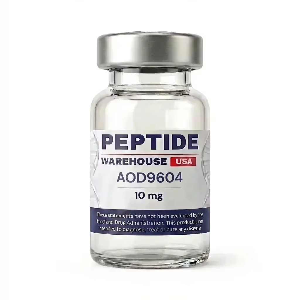
Peptide Warehouse USA offers high-purity research peptides and related compounds for laboratory, analytical, and preclinical applications, with batch documentation such as Certificates of Analysis and stated purity information. Visit Peptide Warehouse USA to explore research materials and supporting documentation, and select products according to your assay requirements, regulatory obligations, and laboratory protocols.
Leave a comment