
No products in the cart.



You're probably here because a peptide sequence is sitting in front of you right now, and a simple question keeps turning into a practical problem. Which residues will make the peptide stay soluble, which ones will push it to clump, and which stretch looks like it wants to bury itself in a membrane?
That question usually leads straight to non polar amino acids. People first meet them as a memorization list in biochemistry, but in research they're a design variable. They affect purification behavior, folding, membrane association, and whether a sequence behaves cleanly or gives you one troubleshooting headache after another.
A good way to think about them is this. The “nonpolar” label is useful, but it isn't the whole story. A residue can look strongly hydrophobic in one setting and behave much more ambiguously in another, especially near membranes, interfaces, or disordered regions. That's where peptide design gets interesting, and where the chemistry starts to matter.
A student opens a peptide file, sees a run of leucines and valines in the middle, and immediately wonders whether the sequence will dissolve at all. A more experienced researcher looks at the same line and asks a different question. Is this segment going to fold inward, stick to another copy of itself, or insert into a lipid environment?
That's why non polar amino acids matter. They often give the earliest clues about solubility, aggregation, folding behavior, and membrane partitioning. If you ignore them, you can spend time and synthesis budget on a peptide that behaves very differently from what the sequence first suggested.
In routine peptide work, nonpolar stretches often correlate with familiar lab outcomes:
The chemistry is simple in spirit. Side chains that don't interact favorably with water often push the molecule toward self-association or toward environments where those groups can be buried.
Practical rule: If a peptide's behavior surprises you, inspect the side chains before you inspect the instrument.
Biochemistry references commonly classify amino acids by the nature of the side chain at neutral pH, separating them into nonpolar, polar uncharged, negatively charged, and positively charged groups, which is exactly why this framework shows up so often in protein structure discussions in this biochemistry overview from LibreTexts.
That classification isn't just for exams. In peptide research, it influences choices about solvent systems, membrane mimics, assay setup, and how much confidence you should place in a predicted conformation. It also shapes how people talk about the benefits of peptides in a research setting, because many useful peptide properties come from balancing hydrophobic and hydrophilic residues rather than maximizing one or the other.
For anyone comparing sequences, screening analogs, or planning a peptide stack for mechanistic studies, nonpolar residues are usually among the first features worth circling.
At the core, an amino acid is called nonpolar because its R group, or side chain, has little tendency to interact strongly with water. It usually carries little to no net charge at neutral pH and usually doesn't offer much hydrogen bonding compared with clearly polar side chains.
That doesn't mean water “hates” the residue in some dramatic way. It means the side chain fits poorly into water's hydrogen-bonding network, so water has to organize around it. That's why nonpolar groups often end up tucked away inside proteins or facing lipid tails instead of solvent.
Oil and vinegar separate even when you shake them hard. The oil isn't attacking the vinegar. It just doesn't mix well with it.
Nonpolar amino-acid side chains behave in a related way. In aqueous environments, they tend to cluster or move toward regions where water contact is reduced. In proteins, that usually means burial. In membranes, it can mean insertion. In short peptides, it can mean aggregation if the rest of the sequence doesn't counterbalance them.
You can spot many non polar amino acids by their side-chain architecture.
One teaching source puts this nicely by emphasizing that very nonpolar side chains are dominated by hydrocarbon content. It explicitly lists Ala, Val, Leu, Ile, Phe, and Met as very non-polar, while placing Gly, Cys, Pro, Trp, and Tyr in a more moderately non-polar category, which is a helpful reminder that nonpolarity isn't always treated as a strict binary in this amino-acid polarity teaching chapter.
The contrast gets clearer when you place them beside other classes.
A lot of confusion disappears once you focus on the side chain rather than the amino acid name. The backbone is shared by all standard amino acids. The R group is what changes the personality.
Many teaching systems split non polar amino acids into aliphatic and aromatic families. That division helps because not all hydrophobes behave the same way. Some are compact hydrocarbon packers. Others bring a ring system that adds bulk, shape, and surface preferences.
A standard reference split places alanine, valine, leucine, isoleucine, and methionine in the aliphatic group, while phenylalanine, tryptophan, and sometimes tyrosine appear in the aromatic group in this biochemistry text on amino-acid classes.
A widely cited set of nonpolar amino acids includes glycine, alanine, valine, leucine, isoleucine, methionine, and proline. Some references expand that list to include phenylalanine, tryptophan, tyrosine, cysteine, or others, which is why it's important to say which grouping method you're using in this Pearson overview of nonpolar amino acids.
That's where most student confusion begins. The “easy” residues are usually Ala, Val, Leu, and Ile. The tricky ones are Gly, Pro, Met, Cys, Trp, and Tyr.
Boundary residues are often the ones that decide whether a peptide acts like a straightforward hydrophobe or a switch that changes behavior with environment.
| Residue | Family | Side-Chain Feature | Hydrophobic Character |
|---|---|---|---|
| Alanine | Aliphatic | Small hydrocarbon methyl group | Clearly nonpolar, modest hydrophobe |
| Valine | Aliphatic | Branched hydrocarbon chain | Strongly hydrophobic |
| Leucine | Aliphatic | Branched hydrocarbon chain | Strongly hydrophobic |
| Isoleucine | Aliphatic | Branched hydrocarbon chain | Very strongly hydrophobic |
| Methionine | Aliphatic | Thioether within hydrocarbon-like chain | Hydrophobic in most protein contexts |
| Proline | Aliphatic-like | Cyclic side chain linked to backbone | Nonpolar but structurally distinctive |
| Glycine | Often grouped with aliphatic/core nonpolar sets | Minimal side chain, just hydrogen | Weakly nonpolar and highly context-dependent |
| Phenylalanine | Aromatic | Phenyl ring | Strong aromatic hydrophobe |
| Tryptophan | Aromatic | Indole ring with N-H | Hydrophobic but interfacially favored in many settings |
| Tyrosine | Aromatic | Phenol ring with hydroxyl | Only marginally hydrophobic compared with cleaner hydrophobes |
Sooner or later, labels like “nonpolar” stop being enough. You need numbers. The classic place many researchers start is the Kyte-Doolittle hydropathy scale, because it gives each amino acid a numeric hydrophobicity value that you can compare across a sequence.
This scale became especially influential after its publication in 1982, and it was widely adopted in protein-sequence analysis, membrane-protein prediction, and structure-function studies. In one commonly used hydrophobicity reference, the strongest hydrophobic values include isoleucine (+4.5), valine (+4.2), leucine (+3.8), phenylalanine (+2.8), cysteine (+2.5), methionine (+1.9), and alanine (+1.8), while polar or charged residues fall much lower, such as aspartic acid (-3.5), glutamic acid (-3.5), and lysine (-3.9) in this hydrophobicity index table.
A residue labeled “nonpolar” can still vary a lot in practical behavior. Isoleucine at +4.5 and alanine at +1.8 don't contribute equally to hydrophobic packing. That difference matters when you're deciding whether a mutation will merely tweak a peptide's behavior or shift it into a different solubility or membrane-binding regime.
It also shows why boundary cases are important. Cysteine at +2.5 can look hydrophobic by one measure, yet many researchers still treat it cautiously because its chemistry can change with oxidation state and local environment.
| Amino Acid | KD Value | Side Chain Class | Design Note |
|---|---|---|---|
| Isoleucine | +4.5 | Nonpolar aliphatic | Strong driver of hydrophobic segments |
| Valine | +4.2 | Nonpolar aliphatic | Common in compact hydrophobic cores |
| Leucine | +3.8 | Nonpolar aliphatic | Frequent in buried and membrane-facing regions |
| Phenylalanine | +2.8 | Aromatic nonpolar | Adds hydrophobic bulk with aromatic character |
| Cysteine | +2.5 | Boundary residue | Often context-sensitive despite positive hydropathy |
| Methionine | +1.9 | Nonpolar aliphatic | Flexible hydrophobe, useful in packing |
| Alanine | +1.8 | Nonpolar aliphatic | Mild hydrophobe, often tolerated in helices |
| Aspartic acid | -3.5 | Negatively charged | Usually disfavors hydrophobic interiors |
| Glutamic acid | -3.5 | Negatively charged | Strongly solvent-oriented in most settings |
| Lysine | -3.9 | Positively charged | Strongly hydrophilic by this scale |
For peptide work, these values are useful in at least three ways:
Other scales exist, and they don't always rank every residue identically. A major review of hydrophobicity measurements notes that values vary by method, but the most nonpolar residues remain remarkably consistent across scales. Leucine, isoleucine, valine, phenylalanine, methionine, and alanine repeatedly appear among the most hydrophobic residues, and later work established a direct water-to-bilayer transfer free-energy scale for all natural side chains in a membrane context in this review and linked research summary.
If a vendor, paper, or software tool reports hydrophobicity, check which scale it's using before you compare outputs.
When proteins fold in water, nonpolar side chains rarely want to stay exposed for long. They tend to move inward, pack together, and form the familiar hydrophobic core that stabilizes many globular proteins.
That process is commonly explained by the hydrophobic effect. In water, exposed nonpolar surfaces are thermodynamically unfavorable at the solvent interface. Folding buries those groups in the protein interior, and that burial lowers free energy by compensating for the entropy loss associated with water reorganization around hydrophobes as described in this PNAS hydrophobic-effect discussion.
A quick visual helps:
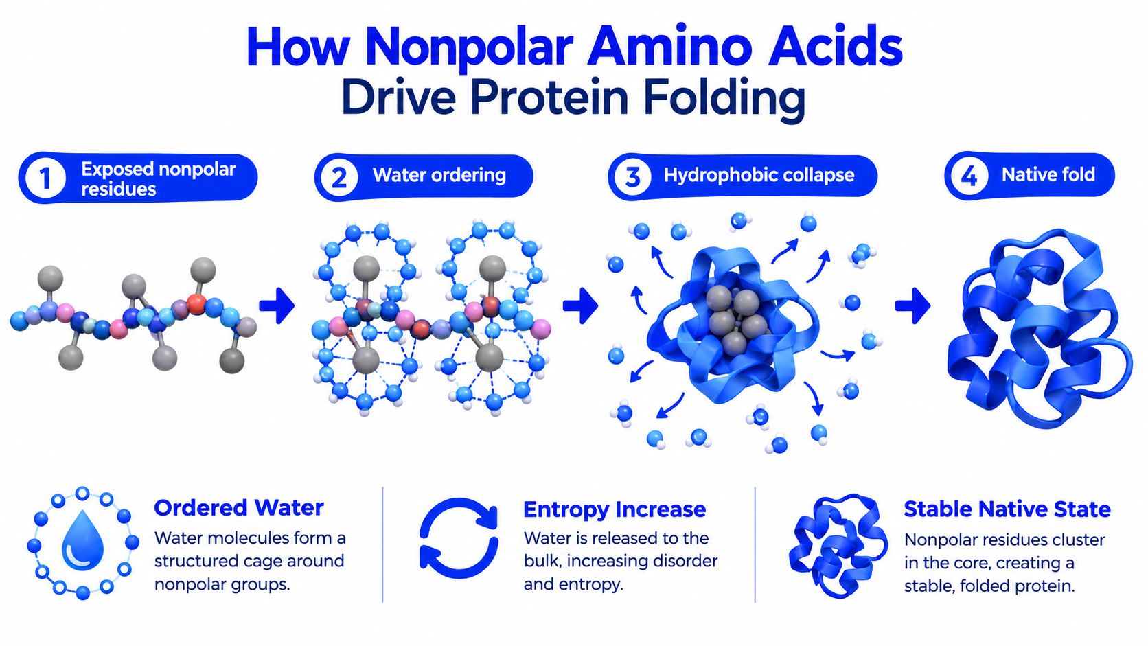
Water likes to hydrogen bond with itself. When a nonpolar side chain interrupts that network, nearby water becomes more ordered. If several hydrophobic side chains cluster together during folding, less nonpolar surface remains exposed, and some of that ordered water is released back into the bulk.
That's why nonpolar residues often act like the inward-facing scaffolding of a protein. They help create a packed interior where side chains fit together like the hidden beams of a structure.
The residues many researchers watch closely in buried regions are the strong hydrophobes such as leucine, isoleucine, valine, and phenylalanine. Aromatic residues can contribute more than bulk hydrophobicity alone because their ring systems can also create distinctive packing interactions.
If you work with structure prediction or conformational screening, it helps to pair this chemistry intuition with modeling tools. PepFlow's 2026 conformation guide gives useful context for thinking about how sequence-level features map onto likely folded or partially folded states.
The folding story is easier to grasp when you watch it described step by step:
A buried hydrophobe usually helps a fold. A buried charge usually asks the structure to pay an energetic price.
That's why swapping a core hydrophobe for a charged residue often destabilizes a protein, even if the sequence still looks similar at a quick glance. In peptide design, it's rarely enough to ask whether a residue is “allowed.” You need to ask whether that residue makes sense in that local environment.
Inside a soluble protein, nonpolar side chains usually point inward. In a membrane protein, many of those same side chains do the opposite. They point outward toward lipid tails because the bilayer core is itself hydrophobic.
That's why transmembrane helices are often enriched in residues such as Leu, Ile, Val, Phe, and Ala. Their side chains are a good chemical match for the membrane interior, while strongly polar or charged residues are generally harder to accommodate in the bilayer core.
Quantitative helix studies make this point very concrete. Nonpolar solvation energy parameters of about -10 cal/(mol·Å^2) for aliphatic surface area and -7 cal/(mol·Å^2) for aromatic surface area show that hydrophobic side-chain surface area strongly favors membrane partitioning and stabilizes helix insertion in this transmembrane-helix analysis.
That gives membrane biophysics a more physical basis than the simple statement that “hydrophobic residues like membranes.” The amount and type of exposed nonpolar surface matter.
Not every membrane-associated segment is fully buried. Many peptides form amphipathic helices, where one face is richer in nonpolar residues and the opposite face carries more polar character. In that arrangement, the peptide can sit at an interface with one side oriented toward lipid tails and the other toward water.
This is also where aromatic residues become especially interesting. A residue may not behave like a pure bilayer-core hydrophobe, but it may still be favored at the membrane interface because its size, polarizability, or ring chemistry suits that transition zone.
Lab takeaway: When a peptide binds lipids but won't fully insert, inspect the pattern of hydrophobic residues, not just the total count.
For researchers working on peptide carriers, membrane-active constructs, or detergent-resistant designs, nonpolar residues are best treated as spatial features. Their placement often matters as much as their identity.
Calling a residue “nonpolar” is useful, but it can mislead you if you treat it like a permanent identity rather than a context-sensitive behavior.
Recent work pushes exactly that point. A 2025 study introduced a dewetting free-energy hydrophobicity scale and explicitly framed amino-acid classification as dependent on entropic and enthalpic amino acid-water interactions rather than a single static label. A related 2025 membrane biophysics review also notes that newer experimental scales distinguish core, interfacial, and aqueous preferences, and that residues often grouped together in textbooks can behave very differently near membranes as summarized in this PubMed-indexed source.
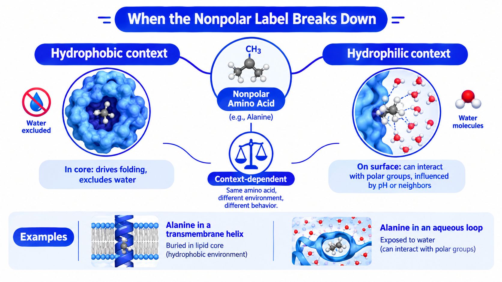
A leucine buried in a protein core and a leucine exposed on a protein surface are chemically the same residue, but they aren't playing the same role. The same goes for tryptophan and tyrosine near a bilayer. They may not fit a simplistic “greasy residue” picture, yet they often behave like effective interfacial anchors.
Cysteine is another good reminder. In one setting, it contributes to a hydrophobic core. In another, it changes character through redox state, coordination, or covalent bonding.
One of the newer research angles concerns liquid-liquid phase separation. A 2025 ACS Biomacromolecules paper found that a single amino-acid model can undergo hydrophobically driven liquid-liquid phase separation depending on pH and ionic strength, which means environmental conditions can flip the apparent behavior of residues that are usually taught as nonpolar. Related 2025 biophysical work also reported that hydrophobicity inferred from partial atomic charge is tied to accessibility and charge distribution in this ACS Biomacromolecules article.
That matters because aggregation, condensate formation, and compact folding aren't the same outcome. A hydrophobic patch might drive any of them depending on sequence pattern and environment.
Treat “nonpolar” as a starting hypothesis, not a verdict.
The most useful hydrophobicity scale is the one that matches the environment your peptide actually sees.
If you remember one thing, remember this. Non polar amino acids are not just a memorized category. They are a working design language for peptides and proteins.
They help you anticipate whether a sequence may bury itself, stick to itself, sit at an interface, or prefer a membrane-like setting. They also help explain why two peptides with similar lengths and charges can behave very differently in purification, storage, or functional assays.
For day-to-day research, this shorter checklist is usually enough:
When you're choosing or refining a research peptide, this framework can support better decisions around:
For peptide and protein researchers, this also connects to broader use cases like biomaterials, membrane-active constructs, and phase-separation models. As hydrophobicity models become more nuanced, the old binary categories still help, but they work best when paired with experimental context and careful sequence reading.
A final habit is worth adopting. Keep a hydropathy scale nearby, but don't let it overrule observation. If your peptide refuses to behave the way the category predicts, the sequence is telling you something about context, not breaking the rules.
Use that tension productively. It's often where the most valuable design insight appears.
If you're sourcing research peptides or related compounds for sequence testing, analytical work, or preclinical studies, Peptide Warehouse USA offers high-purity research products with batch documentation, including COAs and supporting reports that help with traceability and consistency. To explore options for peptide research workflows and learn more about available products, visit Peptide Warehouse USA.
Leave a comment